
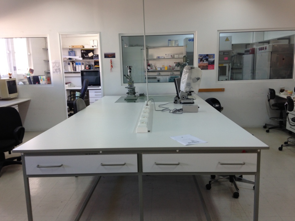
Histology - Embryology lab
Medical School, University of Crete


 Dragana Nikitovic Ph.D., ERT
Associate Professor of
Histology - Embryology
e-mail: nikitovic@uoc.gr
office: +0030 2810 39 4557
lab: +0030 2810 39 4735
Current and completed research projects of the Laboratory of Histology - Embryology
- Study of the synthesis and distribution of GAGs in MG-63 and Saos 2 human osteosarcoma, of high and low metastatic potential cell lines, respectively. The results of the study showed that both human osteosarcoma cell lines synthesize extracellular hyaluronic acid (HA) as well as secreted and cell-attached galactosaminoglycans (GalAGs) and heparan sulfate (HS). Although both cell lines produce significant amounts of proteoglycans (PGs), Saos 2 cells produce HA, GalAGs, and HS in significantly lower amounts than those of MG-63 cells. The effect of genistein on the synthesis of these molecules was also studied. The inhibitory effect of genistein on the synthesis of extracellular secreted and cell-attached GAGs / PGs in Saos 2 cells was found to be dose dependent and most likely exerted through a PTK mechanism. The synthesis of GAGs / PGs by MG-63 cells in the presence of genistein depends on their type and distribution, suggesting a more complex mechanism that regulates the synthesis of PGs.
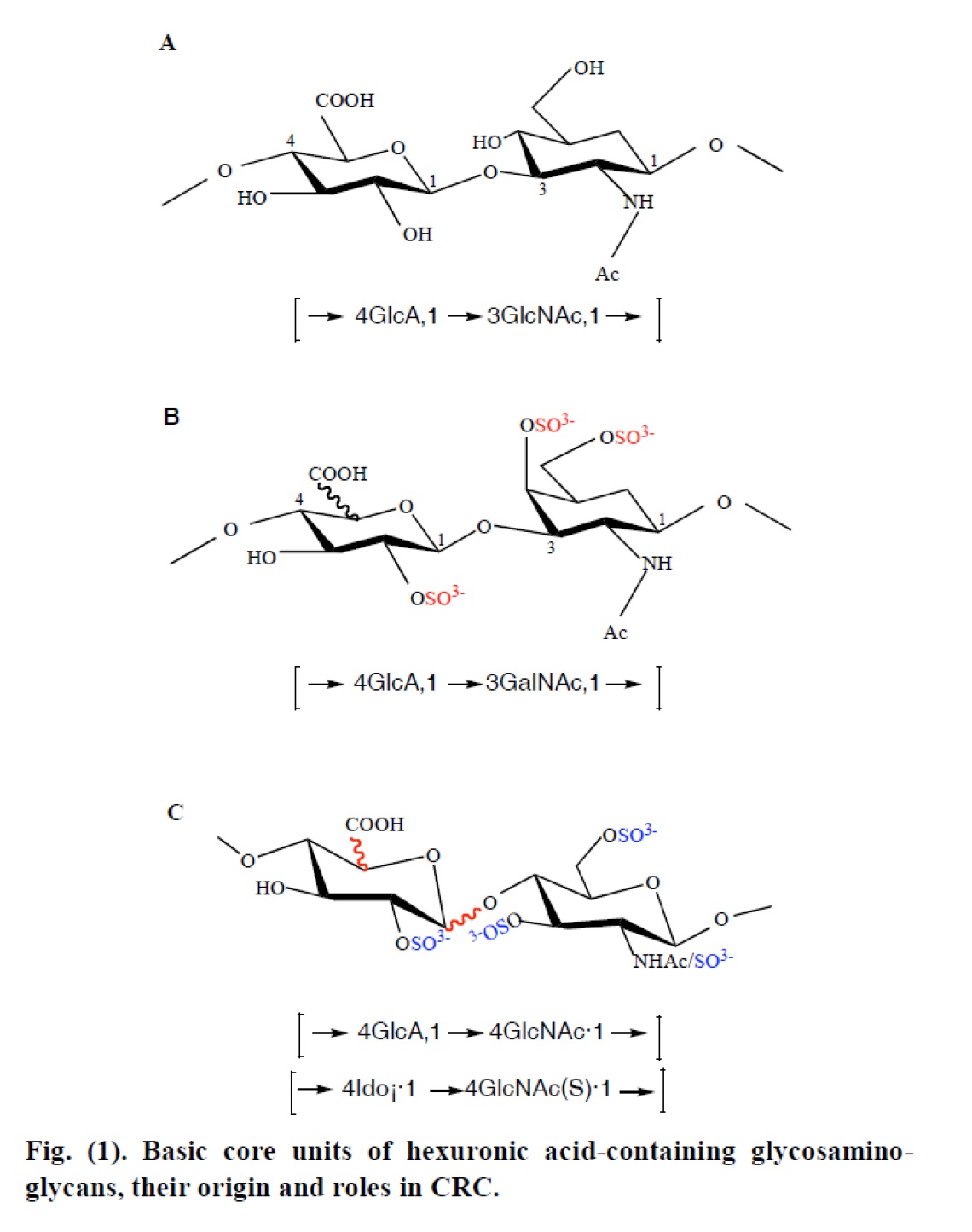
- Assessing the effect of TGF-β2, bFGF and PDGF-BB on the synthesis and distribution of GAGs / PGs by the two osteosarcoma cell lines. The results showed that the action of growth factors differs between the two cell lines and that the regulation of GAGs synthesis depends on their type and distribution. Modifications in the structural composition of the GAGs / PGs constituents of the extracellular matrix can have important effects on cell proliferation and / or differentiation. In human osteoblasts, as well as in the MG-63 and Saos 2 osteosarcoma cell lines, the expression of major GAGs types, e.g. chondroitin sulfate (CSA), dermatan sulfate (DS) and heparin (Hep) was studied as well as their effect on the growth of these cells. The results showed that extracellular matrix GAGs are macromolecules that affect the cell growth of malignant and normal osteoblastic cells in a dose-dependent manner. These effect are closely related to the detailed chemical structure of GAGs, such as the presence of L-iduronic acid and the degree of their sulfation. In addition, taking into account that TGF-β2, bFGF and PDGF-BB are important regulators of extracellular matrix macromolecule expression, the effect of these growth factors on the expression of the large chondroitin sulfate PG, Versican, isoforms, as well as on the synthesis of HA by MG-63 osteosarcoma cells and normal human periodontal ligament osteoblasts (hPDL) was examined. The results showed that TGF-β2 enhanced the expression of Versican and HA by human osteosarcoma cells, whereas PDGF-BB exerted its main effects on the expression of HAS2 isoform and HA biosynthesis by osteoblasts. In conclusion, TGF-β2 is suggested to have a role in the metastatic potential of human osteosarcoma cells.
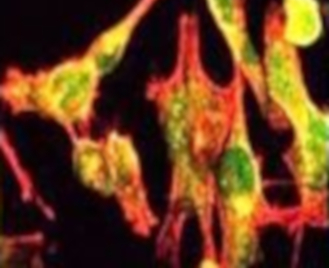
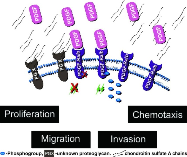
- Study of chondroitin sulfate A chains (CSA) in the regulation of proliferation, adhesion, migration and chemotaxis of both fibrosarcoma cells and normal fibroblasts. Specifically, it was demonstrated that CSA can enhance the mitogenic activity of platelet-derived growth factor in fibrosarcoma cells utilizing a pathway which involves tyrosine kinases whereas; CSA prevents platelet derived growth factor-mediated phosphorylation of PDGF-R beta in normal human fibroblasts severely impairing mitogenic responses. CS chains were found to upregulate fibrosarcoma cell motility through the MAPK pathway, specifically through JNK, whereas CS-induced migration was found to require tyrosine kinase dependent pathways.
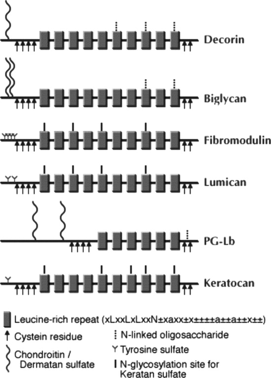
- Study of the role of proteoglycans and glycosaminoglycans on the basal and FGF-2-induced proliferation, adhesion and migration of melanoma cells and normal melanocytes focusing on Erk1/2 nad FAK downstream signaling pathways. In more detail, it was demonstrated that chondroitin/dermatan sulfate-containing proteoglycans, likely in cooperation with heparan sulfate, participate in metastatic melanoma cell FGF-2-induced mitogenic response. Moreover, it was shown that lumican, a small leucine-rich proteoglycan substituted with keratan sulfate chains is expressed and secreted by human melanoma cells and not bu normal melanocytes.
- Study of the putative role of proteoglycans / glycosaminoglycans (PGs / GAGs) on colon cancer cell biological functions. It was demonstrated that PGs / GAGs production of these cells was regulated by both estrogen receptor and protein tyrosine kinase signaling pathways. Importantly, this study introduces a novel role for heparin, as it was shown that this GAG finely modulates the expression of genes crucial to cell cycle regulation through specific activation of p38 MAP kinase to stimulate colon cancer cell growth.
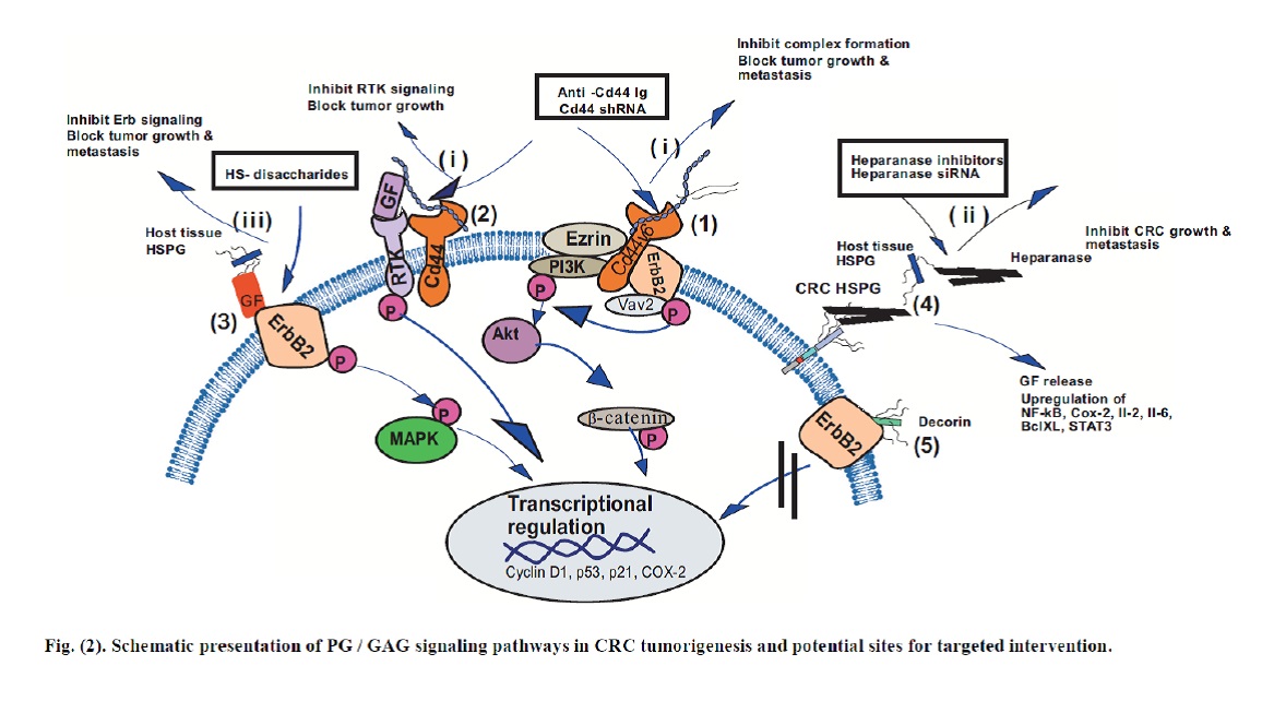
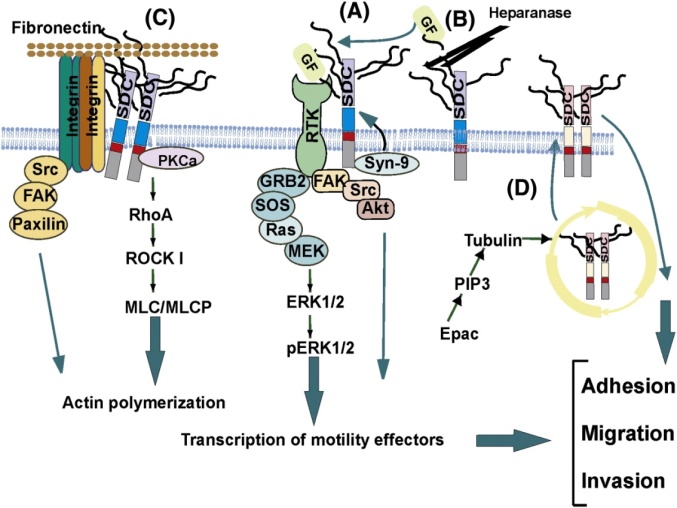
- Study of the role of heparin and heparin sulfate proteoglycans on melanoma cell biological functions. Specifically, it was demonstrated that the action of high molecular weight heparin in the regulation of melanoma cell adhesion and migration involves a p53/FAK/signaling pathway. Moreover, it was shown that fibroblast growth factor-2 modulates melanoma adhesion and migration through a syndecan-4-dependent mechanism. Furthermore, it was demonstrated that low molecular weight heparin through the downregulation of pPKCa and redistribution to nuclear region attenuates JNK activation, which in turn induces cytoskeleton changes correlated to M5 cell decreased adhesion/migration.
- Osteosarcoma is characterized by the deposition of abundant, partially mineralized extracellular matrix (ECM). In this study it was examined how changes in the ECM affect osteosarcoma migration. Specifically, it was shown that the changes of HA metabolism induced by PTH (1-34) and PTH (7-84) peptides in moderately MG-63 and well-differentiated Saos 2 osteosarcoma cell lines, are correlated to their migration capabilities. The obtained data suggests that there is a regulatory effect of PTH (1-34), in an administration mode-dependent manner, on HA metabolism that is essential for osteosarcoma cell migration. In parallel, it was shown that parathyroid hormone affects the fibroblast growth factor-proteoglycan signaling axis to regulate osteosarcoma cell migration.
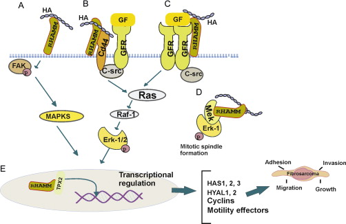
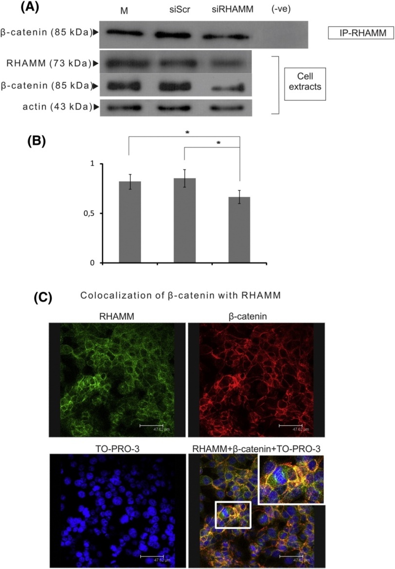
- This study focused on the role of hyaluronan (HA) and its CD44 and RHAMM respective receptors in the regulation of fibrosarcoma biological functions. Importantly, fibrosarcomas were previously shown to have a high content and turnover of ECM components including HA, proteoglycans, collagens, fibronectin, and laminin. The results suggest that RHAMM/HA interaction regulates fibrosarcoma cell adhesion via the activation of FAK and ERK1/2 signaling pathways. Furthermore, the study suggests that RHAMM is a novel β-catenin intracellular binding partner, protecting from degradation and supporting the nuclear translocation of this key cellular mediator, which results in c-myc activation and enhanced fibrosarcoma cell growth.
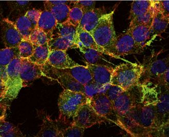
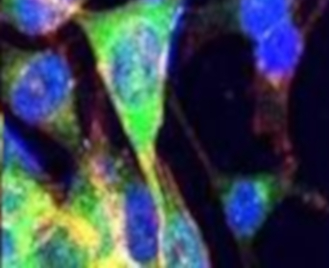
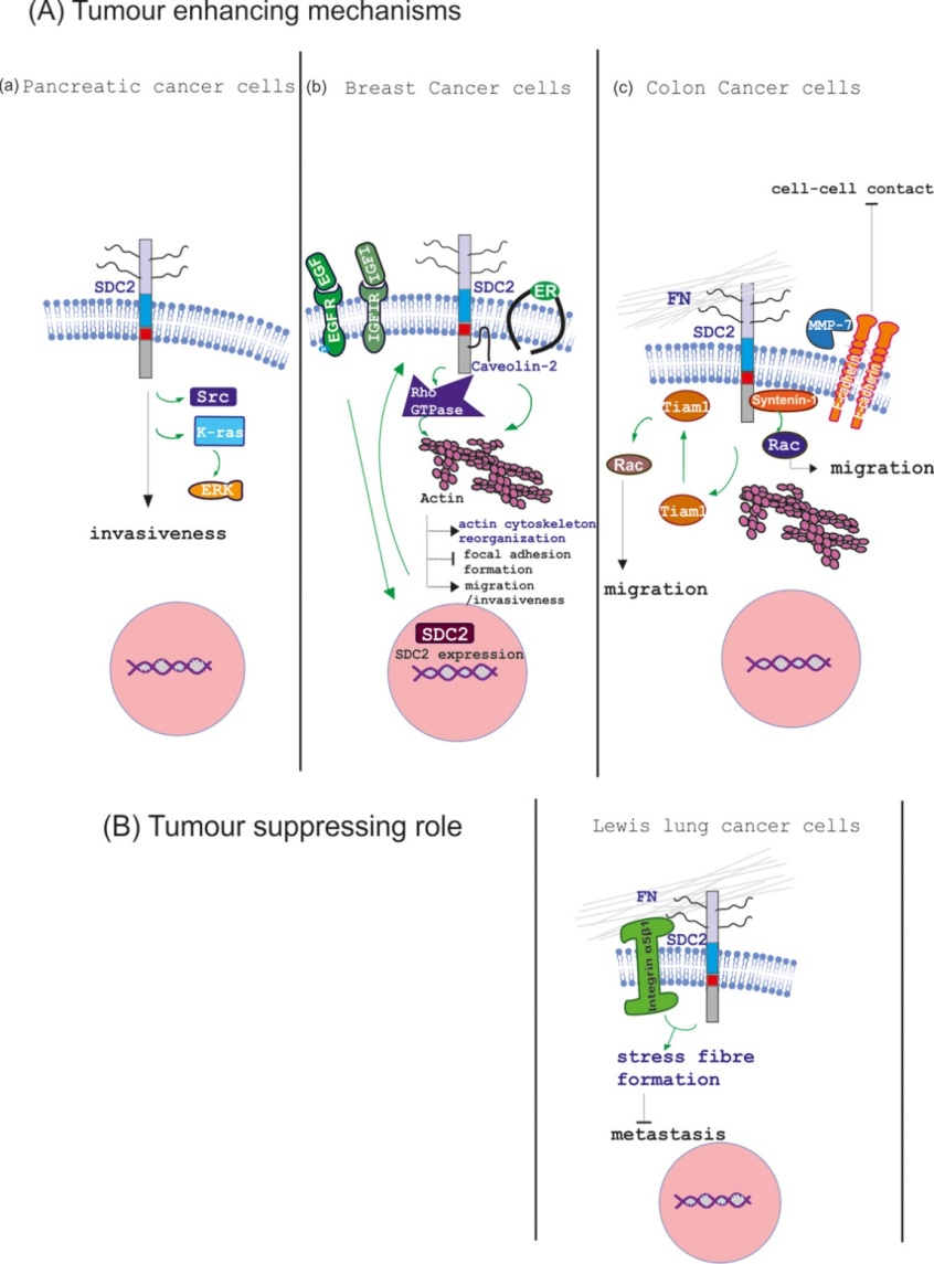
- The study focused on the mechanisms involved in human fibrosarcoma biological functions. In this study, the ability of TGFβ2 to enhance the adhesion of HT1080 cells to fibronectin (FN) by a mechanism involving syndecan 2 (SDC2) and Smad2, was shown. Furthermore, it was demonstrated that the heparin sulfate (HS) chains affect the TGFβ-induced adhesion of HT1080 cells. This study demonstrates for the first time the coexistence of SDC2 with IGF-IR, thereby highlighting SDC2 as a potential co-receptor for IGF-I. Furthermore, downstream activation of ERK1 / 2, which is known to regulate the transcription of essential proteins regulating cell behavior, was demonstrated. At the same time, this study confirmed that ezrin coexists with SDC2 as well as actin at the microtubules of HT1080 cells, thereby partially regulating cell motility and aggressive behavior.
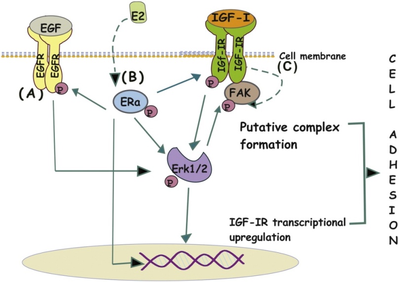
- Study of the role of EGF and IGF growth factors and their EGFR, HER-2 and IGFR respective receptors in breast cancer cells’ functions as well as of the contribution of extracellular macromolecules. This study focused on the interactions of the E2 and IGF-I / EGF molecular signaling pathways, in the regulation of breast cancer cell adhesion as well as on potential intracellular mediators. Fibronectin, an abundant component of the breast cancer cells’ extracellular matrix, is a high molecular weight glycoprotein, which has been described as an aggression marker in the biopathology of breast cancer. In the present study, it has been shown that both IGF-I / EGF and E2 significantly enhance the adhesion of MCF-7 breast cancer cells to the fibronectin substrate. In addition, the main IGF-I receptor, IGF-IR, is required for the induction of adhesion by IGF-I / EGF and E2. Inhibition of Erk1 / 2 inhibits IGF-I- / EGF- / E2- dependent adhesion of MCF-7 cells, demonstrating that Erk1 / 2 is an important intracellular signal mediator of above growth factors for the respective cell function. Administration of IGF-I and EGF induces the reorganization of MCF-7 breast cancer cells’ actin cytoskeleton fibrils. This reorganization appears to be modulated by the inhibition of Erk1 / 2, which is the major cellular mediator of IGF-I and EGF. Interestingly, the administration of IGF-I induces the co-localization of IGF-IR and FAK, which is mainly located to the cell membranes of MCF-7 cells. These data demonstrate that IGF-IR is a convergence point for IGF- / EGF- and E2- dependent cell adhesion onto fibronectin substrate.
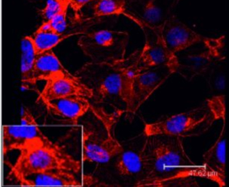
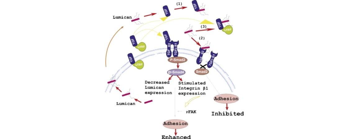
- Study of the effect of small leucine-rich small proteoglycans (SLRPs) on the biological functions of mesenchymal tumors focusing on osteosarcoma and chondrosarcoma. This study has shown that SLRP lumican is expressed by human osteosarcoma cells in a manner dependent on the stage of differentiation of these cells. Lumican was shown to affect the proliferation, migration, and chemotactic response to fibronectin of human osteosarcoma cells, partially, through the TGF-B2 / Smad2 axis. In addition, it was found that the tumor-suppressing SLRP, decorin, did not induce the expression of p21WAF-1, nor did it induce prolonged recycling and inactivation of EGFR. In contrast, EGFR appeared to be overexpressed and to remain in the state of prolonged phosphorylated. In addition, it has been observed that elevated levels of EGFR do not require EGFR activation, suggesting that stabilization of these receptors can be achieved through the interactions of extracellular decorin and EGFR. It is concluded that MG-63 cells are resistant to growth inhibition induced by decorin.
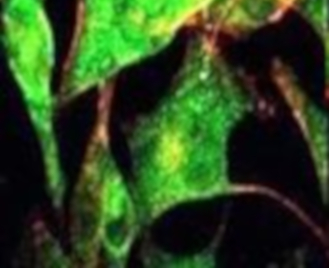
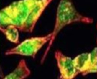
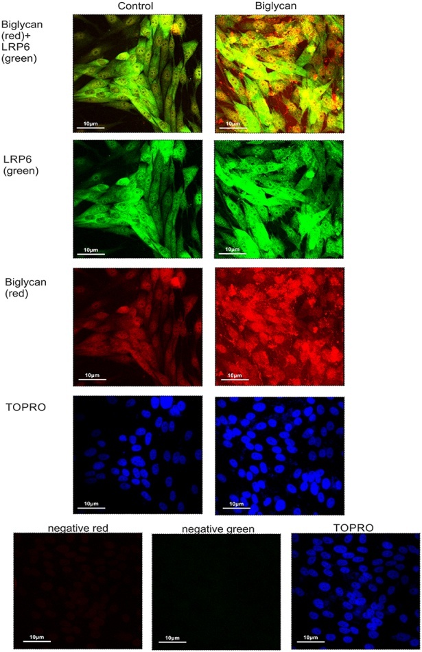
- We also identified a novel mechanism whereby SLRP biglycan through an LRP6 / β-catenin / IGF-IR signaling axis enhances osteosarcoma cell growth. The exact mechanism of the action of biglycan on MG63 cells’ growth is still being studied. We are focus on identifying the components of the IGF-IR / β-catenin intracellular co-localization complex as well as upstream potential regulators. The significance of this mechanism will be explored in other osteosarcoma cell lines, including Saos2, as well as in other mesenchymal-derived tumors, such as HTB94 chondrosarcoma cells. At the same time, we are studying the effects of biglycan and lumican on the cellular functions of chondrosarcoma cells in combination with the growth factors TGFβ2 and IGF-I. We have shown that HTB94 chondrosarcoma cells express lumican which regulates the proliferation of these cells through an IGF-IR / Erk1,2 / p53 signaling axis.
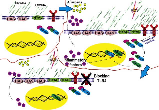
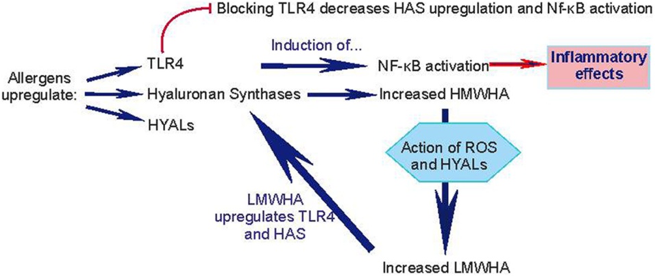
- Study of the role of hyaluronic acid (HA) on keratinocyte activation by allergens. Contact of the skin with unwanted industrial or environmental factors can result in contact dermatitis (ACD) of either allergic or irritating etiology. During inflammation or injury, ECM molecules are released, including fragments of low molecular weight hyaluronic acid (HA) that act as damage-associated molecular patterns (DAMPs) or "danger signals", that is, endogenous molecules that cause and enhance inflammation. Our group showed that the degradation of HA in keratinocytes is altered after incubation with allergens such as DNCB and PPD. In addition, low molecular weight hyaluronan (LWMHA) was found to induce interleukin-18 production, an established indicator of keratinocyte activation. We showed in NCTC2544 human keratinocytes that the contact allergens, PPD and DNCB, induce in a concentration-dependent manner a significant increase of TLR-4 expression. In conclusion, keratinocyte activation is partially perpetrated through the low molecular weight hyaluronic acid / TLR4 / NF-κB signaling axis.
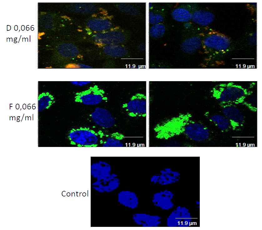
- Study of the interactions of nanoparticles and blood-brain barrier: evaluation of immunological activation and the role of the subendothelial extracellular matrix. The blood-brain barrier (BBB) is a functional cellular barrier between tissue and endothelial cells. The purpose of this study is to evaluate the possible immunological activation of endothelial cells by administering a series of PVP nanoparticles separately or as carrier-drug complexes. The expression of endothelial dysfunction biomarkers and the affinity of peripheral blood leukocytes to the endothelial cell layer were examined. As the ECM is strongly remodelled in inflammation, the potential effects of nanoparticles on the expression of subendothelial ECM components including collagen IV, fibrinectin, laminin, heparin, and heparin are carried out in our laboratory.

INNOvation with GLYcans
Faculty of Medicine, University of Crete
Hybrid Symposium
27-29 September
Heraklion, Greece & Zoom Platform
Poster, Timetable
Special Issue
"Exploring the Multifaceted Roles of Glycosaminoglycans (GAGs) - New Advances and Further Challenges"
Editors
Dr. Dragana Nikitovic
Dr. Serge Perez
Website
Special Issue
"The Role of Extracellular Matrix in Cancer Development and Progression"
Editors
Professor George Tzanakakis
Professor Dragana Nikitovic
Website
